Brain Evolution & Myofascial Correspondence
From Smooth Brains to Complex Cortices: How Peripheral Myofascial Nerve Networks Sculpt Cortical Folding Patterns Through 66 Million Years of Mammalian Evolution
Core Thesis
Peripheral myofascial nerve connection complexity directly corresponds to cortical folding complexity, with peripheral myofascia serving as intersections between body fascia groups and brain folds serving as intersections of their corresponding brain regions.
Challenging the Current Paradigm: Current neuroscience explains cortical folding through mechanical tension during development - where the outer cortical layer grows faster than the underlying white matter, creating physical tension that causes the brain surface to buckle and fold into gyri and sulci. This thesis proposes that this developmental mechanical tension is not random, but is itself precisely guided by the peripheral myofascial nerve network architecture.
The Direct Correspondence: Each peripheral myofascial intersection - where fascial planes converge and create complex proprioceptive nerve networks - has a corresponding cortical fold intersection where multiple brain regions converge. The complexity of nerve connections at peripheral myofascial junctions directly determines the depth and complexity of the corresponding cortical sulci and gyri. This creates a precise anatomical mapping between body and brain architecture.
The Revolutionary Insight: The mechanical tension that physically sculpts cortical folds during brain development is directed by the peripheral myofascial nerve network. Each gyrus and sulcus represents not just additional surface area, but the brain's architectural adaptation to process and integrate with specific myofascial intersection points. The brain's folding pattern is literally a neural map of the body's fascial intersection complexity - making cortical folding the central nervous system's mirror of the peripheral tensegrity network.
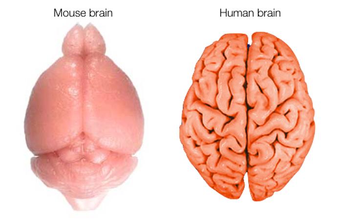
Early Mammals (Rodentia)
Movement Complexity Profile
- Locomotion: Quadrupedal scurrying in 2D plane with low center of gravity
- Manipulation: Basic paw use for holding food, no fine motor control
- Behavioral Repertoire: Instinctual, repetitive patterns for survival tasks
Brain Structure
- Small brain with almost no gyrification
- Represents baseline mammalian brain structure
- Minimal cortical surface area
Lagomorpha
Movement Complexity Profile
- Locomotion: Explosive hopping and bounding requiring whole-body coordination
- Evasive Maneuvers: Rapid, unpredictable directional changes to evade predators
- Shock Absorption: Complex myofascial system for landing impacts
Brain Structure
- Mostly smooth but with beginning of slight indentations
- Enhanced cerebellum for coordination
- Step up from basic rodent structure
Carnivora
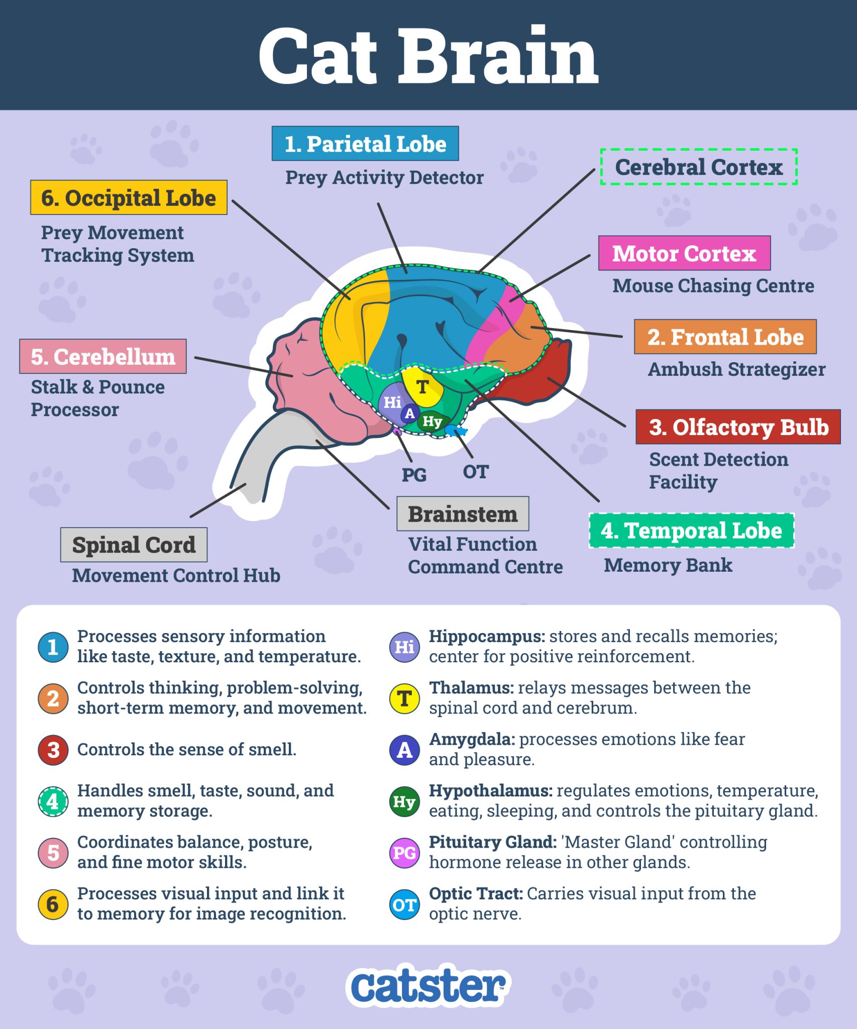
Movement Complexity Profile
- Predatory Dynamics: Stalking, sprinting, leaping, climbing, mid-air body twisting
- Sensorimotor Integration: Rapid processing of visual, auditory, and proprioceptive data
- Fine Control: Delicate paw manipulation for prey handling
Brain Structure
- Numerous distinct gyri and sulci
- Significant leap in cortical surface area
- Enhanced motor and sensory processing regions
Perissodactyla
Movement Complexity Profile
- Specialized Locomotion: High-speed, rhythmic movement over varied terrain
- Biomechanical Complexity: Shock absorption and energy storage through fascial systems
- Herd Dynamics: Coordinated group movement requiring social motor integration
Brain Structure
- Large and densely folded brain
- More complex than carnivore brains
- Specialized for rhythmic motor control
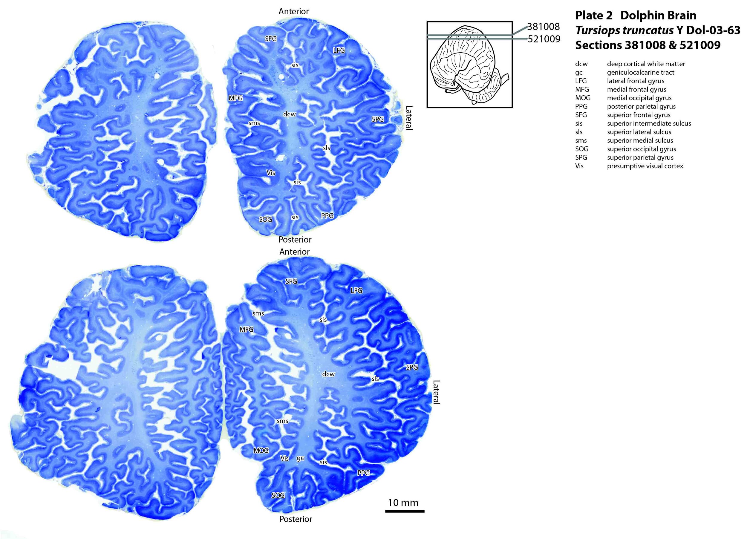
Cetacea
Movement Complexity Profile
- Six Degrees of Freedom: Movement and rotation on all three axes in 3D fluid environment
- Echolocation: Real-time sound emission, echo processing, and spatial navigation
- Hydrodynamics: Precise control of flukes and flippers for graceful maneuvering
Brain Structure
- One of the most convoluted brains in nature
- Surface area rivals and sometimes exceeds humans
- Extreme gyrification for 3D navigation
Early Primates
Movement Complexity Profile
- Arboreal Locomotion: Generalized arboreal quadruped with grasping hands and feet
- 3D Navigation: Basic navigation in branch-filled environment
- Distance Judgment: Leaping between branches requiring spatial assessment
Brain Structure
- Small, relatively lissencephalic brain
- Notable expansion in visual processing areas
- Enhanced compared to ground-dwelling contemporaries
Early Apes

Movement Complexity Profile
- Enhanced Flexibility: Increased mobility in shoulder, elbow, and wrist joints
- Varied Arboreal Movement: More climbing, clambering, and reaching than predecessors
- Generalized Arborealism: Not specialized swingers but more flexible than early primates
Brain Structure
- Larger brains with clear increase in gyrification
- Prominent visual cortex development
- Enhanced frontal and parietal lobes
Great Apes
Movement Complexity Profile
- Knuckle-Walking: Development of specialized terrestrial locomotion
- Dual-Mode Locomotion: Proficiency in both arboreal and terrestrial environments
- Tool Use: Basic tool use for food processing and insect probing
Brain Structure
- More complex motor cortex
- Significantly larger cerebellum
- Enhanced coordination centers
Early Hominins
Movement Complexity Profile
- Facultative Bipedalism: Could walk upright but retained climbing adaptations
- Dual-Mode Existence: Managing both bipedal and arboreal locomotion
- Balance Challenge: Constant adjustment required for unstable bipedal stance
Brain Structure
- Brain size similar to chimpanzees (350-400cc)
- Forward-positioned foramen magnum
- Reorganized brain base and cerebellum
Australopithecines
Movement Complexity Profile
- Committed Bipedalism: Lost grasping foot, dedicated to upright walking
- Efficient Locomotion: Perfected bipedal gait for long-distance travel
- Retained Climbing: Long arms and curved fingers for occasional arboreal activity
Brain Structure
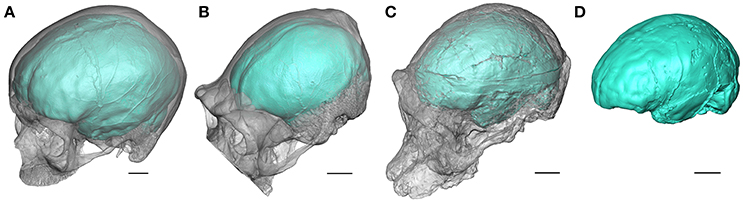
- Increased brain size (400-550cc)
- Backward shift of lunate sulcus
- Expanding parietal cortex for spatial integration
Homo habilis
Movement Complexity Profile
- Systematic Tool Making: Creation of Oldowan stone tools requiring precise motor planning
- Hand-Eye Coordination: Striking stones at exact angles and force
- Sequential Planning: Multi-step processes from material selection to tool completion
Brain Structure
- Significant jump in brain size (550-700cc)
- First clear evidence of expanded Broca's area
- Enhanced motor planning regions
Homo erectus
Movement Complexity Profile
- Long-Distance Migration: First hominin to leave Africa, expert endurance locomotion
- Advanced Tool Technology: Acheulean hand-axes requiring complex mental templates
- Cooperative Hunting: Group coordination for large game hunting
- Fire Control: Complex motor sequences for fire management
Brain Structure
- Major leap in brain size (800-1200cc)
- Significant expansion in frontal and parietal lobes
- More modern-looking, highly folded structure
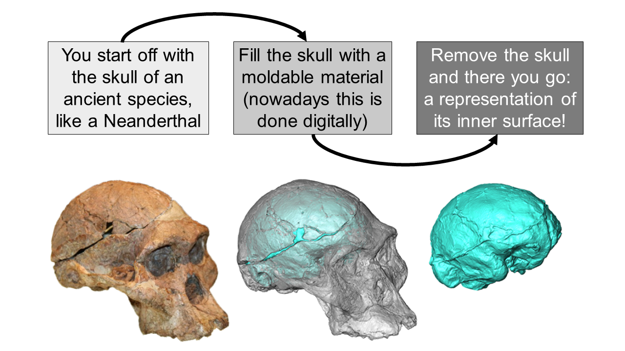
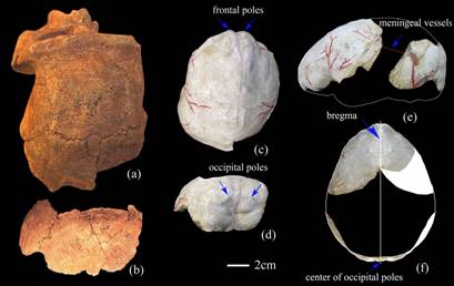
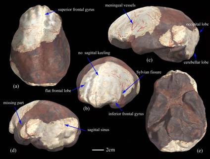
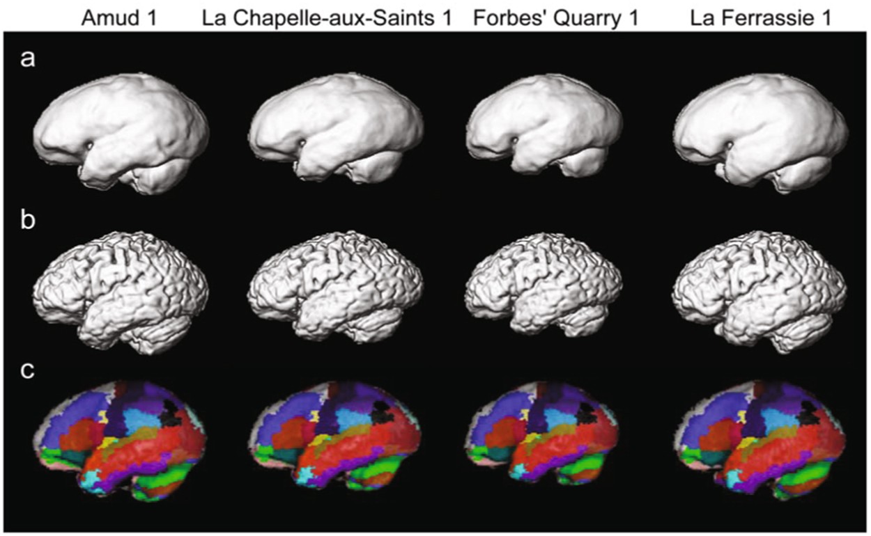
Archaic Humans
Movement Complexity Profile
- Expert Hunting: Sophisticated toolkits for large game hunting in harsh environments
- Complex Manufacturing: Mousterian tools, wooden spears, tailored clothing
- Symbolic Behavior: Use of pigments, burial practices, abstract motor actions
- Environmental Adaptation: Survival in Ice Age conditions requiring complex motor skills
Brain Structure
- Brain size reached modern peak (1200-1750cc)
- Highly gyrified with large frontal, parietal, and occipital lobes
- Neanderthals had larger brains than modern humans on average
Homo sapiens
Movement Complexity Profile
- Projectile Technology: Spear-throwers, bows requiring incredible motor skill and physics intuition
- Art and Music: Cave paintings, beads, instruments representing pinnacle of fine motor control
- Language: Most complex motor sequencing task - coordinating dozens of muscles for infinite meaningful utterances
- Abstract Planning: Ability to invent and perfect entirely novel movement sequences
Brain Structure
- Unique globular shape with enhanced connectivity
- Larger parietal and cerebellar regions
- Extreme gyrification for maximum processing power